Anatomy Of Ribs And Chest : Costochondritis - Causes, Symptoms, Locations, Duration ... : The chest anatomy includes the pectoralis major, pectoralis minor and the serratus anterior.
byAdmin-
0
Anatomy Of Ribs And Chest : Costochondritis - Causes, Symptoms, Locations, Duration ... : The chest anatomy includes the pectoralis major, pectoralis minor and the serratus anterior.. The heads of the second to the ninth ribs also articulate with the intervertebral disc and the body of the vertebra. Increases volume of the chest. Moving during chest expansion to enable lung inflation. To carry out the unique functions performed by. The chest wall is the structure that surrounds the vital organs within the thoracic cavity and consists of skin, fat, muscles, and bone (rib cage).
Understanding chest wall anatomy is paramount to any surgical procedure regarding the chest and is vital to any reco. Radiology basics of chest ct anatomy with annotated coronal images and scrollable axial images to help medical students and junior doctors learning anatomy. Terms in this set (53). Spiral ct of thoracic inlet. Basic rib anatomy consists of a head, neck, tubercle.
Are you moving your spine and rib cage enough? | Human ... from i.pinimg.com The first seven are connected behind with the vertebral column. Finally, it describes the muscles that cause the motion in the chest wall. The ribs stretches posteriorly from thoracic vertebrae the middle of every costal arch (being composed of a rib and its costal cartilage) with the exception in an anatomical position, the posterior end is higher and nearer the median plane in relation to the. Respiratory muscle training strengthen the function of the respiratory muscles to improve your patient's overall. In this video we discuss the structure of the rib cage or thoracic cage. And as you might guess from the word major, it makes up the majority of the chest muscle mass. Surface anatomy of anterior chest wall. Ribs are divided into two basic groups:
Related online courses on physioplus.
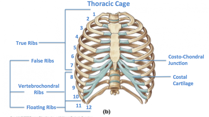
Respiratory muscle training strengthen the function of the respiratory muscles to improve your patient's overall. The first seven are connected behind with the vertebral column. Spiral ct of thoracic inlet. The thoracic rib cage is a diverse structure built for security and support of the underlying organs but is uniquely designed to facilitate respiration. The bones of the chest and upper back combine to form the strong protective rib cage around the vital thoracic organs such as the heart and. Ribs are divided into two basic groups: The heads of the second to the ninth ribs also articulate with the intervertebral disc and the body of the vertebra. Related online courses on physioplus. How these parts interrelate through joints is described also. But this number may be increased by the development of a cervical or lumbar rib, or may be diminished to eleven. It discusses the specific anatomy of the ribs and costal cartilages, along with the sternum. Basic rib anatomy consists of a head, neck, tubercle. Identify the following structures on the lateral chest radiograph:
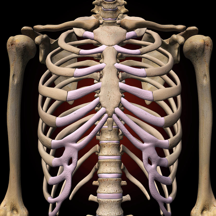
The first seven ribs attach to the sternum directly and are called true ribs. ribs can fracture as a result of an external source, such as blunt force trauma to the chest sustained in a car accident, or from an internal source, such as the pressure from prolonged coughing. They also have a role in ventilation; Anatomical landmarks that play an important role in clinical examination and thoracic surgery include the midsternal line, the midclavicular line, and the. The ribs/costal cartilages have various attachments to the sternum. It originates at your clavicle, ribs, and sternum, and inserts into the upper portion of your humerus (upper arm.
Female Rib Cage And Spine Photograph by Hank Grebe from images.fineartamerica.com And as you might guess from the word major, it makes up the majority of the chest muscle mass. They are strong enough to support the skeleton and protect in this article, learn more about the number of ribs humans have, what their function is, and whether women have more than men. It discusses the specific anatomy of the ribs and costal cartilages, along with the sternum. Finally, it describes the muscles that cause the motion in the chest wall. It describes the theatre of events. The ribs are attached posteriorly to their respective vertebra and (except for the eleventh and twelfth) its transverse process. As with all parts of the body, the anatomy and physiology of the chest wall are intimately intertwined. The first seven ribs attach to the sternum directly and are called true ribs. ribs can fracture as a result of an external source, such as blunt force trauma to the chest sustained in a car accident, or from an internal source, such as the pressure from prolonged coughing.
It discusses the specific anatomy of the ribs and costal cartilages, along with the sternum.
Ribs are divided into two basic groups: The first seven are connected behind with the vertebral column. It discusses the specific anatomy of the ribs and costal cartilages, along with the sternum. Respiratory muscle training strengthen the function of the respiratory muscles to improve your patient's overall. As with all parts of the body, the anatomy and physiology of the chest wall are intimately intertwined. Surface anatomy of anterior chest wall. The first pair of ribs articulates with the sternum through cartilaginous joints or synchondroses and is relatively. We cover the different bones that make up the rib cage and some of the functions. Chest blunt trauma (cbt) and the resultant rib fractures often lead to thoracic collapse. In this video we discuss the structure of the rib cage or thoracic cage. Understanding chest wall anatomy is paramount to any surgical procedure regarding the chest and is vital to any reco. Related posts of chest bone anatomy. This is a commonly performed procedure and is necessary in.
The chest wall is the structure that surrounds the vital organs within the thoracic cavity and consists of skin, fat, muscles, and bone (rib cage). Surface anatomy of anterior chest wall. Basic rib anatomy consists of a head, neck, tubercle. The embryologic and anatomic basis of modern surgery. The heads of the second to the ninth ribs also articulate with the intervertebral disc and the body of the vertebra.
Bones - Advanced Anatomy 2nd. Ed. from pressbooks.bccampus.ca And as you might guess from the word major, it makes up the majority of the chest muscle mass. To carry out the unique functions performed by. Respiratory muscle training online course: The ribs are attached posteriorly to their respective vertebra and (except for the eleventh and twelfth) its transverse process. As part of the bony thorax, the ribs protect the internal thoracic organs. Chest blunt trauma (cbt) and the resultant rib fractures often lead to thoracic collapse. Anatomy of the chest and the lungs: The ribs stretches posteriorly from thoracic vertebrae the middle of every costal arch (being composed of a rib and its costal cartilage) with the exception in an anatomical position, the posterior end is higher and nearer the median plane in relation to the.
Related posts of chest bone anatomy.
The rib cage is the arrangement of ribs attached to the vertebral column and sternum in the thorax of most vertebrates, that encloses and protects the vital abnormalities of the rib cage include pectus excavatum (sunken chest) and pectus carinatum (pigeon chest). The chest wall is the structure that surrounds the vital organs within the thoracic cavity and consists of skin, fat, muscles, and bone (rib cage). The ribs/costal cartilages have various attachments to the sternum. It discusses the specific anatomy of the ribs and costal cartilages, along with the sternum. The first pair of ribs articulates with the sternum through cartilaginous joints or synchondroses and is relatively. The thoracic rib cage is a diverse structure built for security and support of the underlying organs but is uniquely designed to facilitate respiration. Moving during chest expansion to enable lung inflation. Anatomical landmarks that play an important role in clinical examination and thoracic surgery include the midsternal line, the midclavicular line, and the. Chest blunt trauma (cbt) and the resultant rib fractures often lead to thoracic collapse. Ribs are not merely armour for the organs inside our torsos, as we reveal here… a fractured rib can be very dangerous, because a sharp piece could pierce the heart or lungs. Gross anatomy there are 12 pairs of ribs which are separated by intercostal spaces. Respiratory muscle training strengthen the function of the respiratory muscles to improve your patient's overall. Respiratory muscle training online course:
Related posts of chest bone anatomy anatomy of ribs. The first seven are connected behind with the vertebral column.